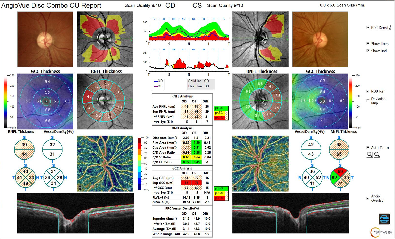
En Face OCT Superior to Red-free Imaging for RNFL Defects
4.7
$ 23.00
In stock
(704)
Product Description
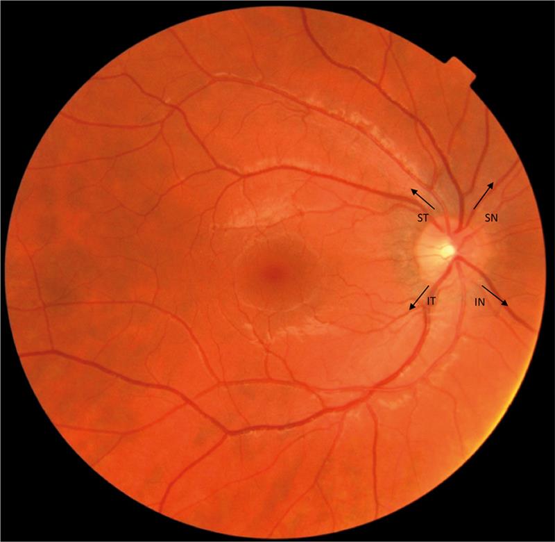
CET: Blood supply to the retina
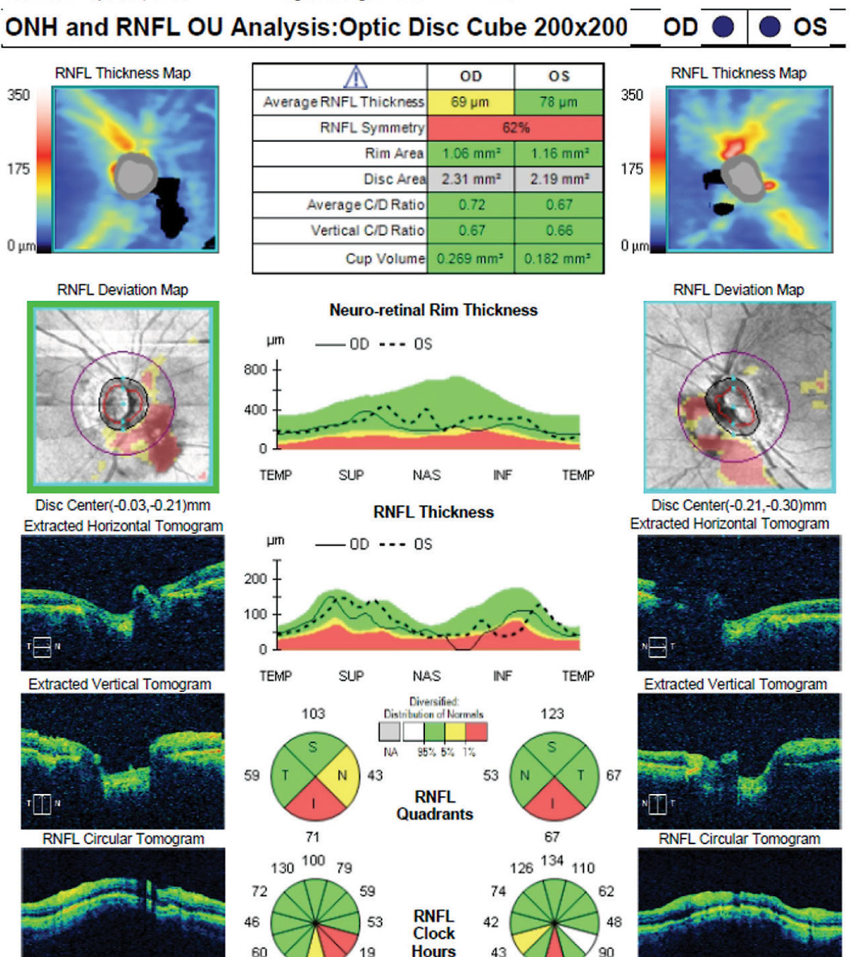
Lesson: Maximizing OCT in the Diagnosis and Management of Glaucoma

Mistakes not to make in glaucoma management - Optometry Australia

Macular imaging by optical coherence tomography in the diagnosis and management of glaucoma

Parapapillary deep‐layer microvasculature dropout is only found near the retinal nerve fibre layer defect location in open‐angle glaucoma - Son - 2022 - Acta Ophthalmologica - Wiley Online Library

Comparison of localised nerve fibre layer defects in normal tension glaucoma and primary open angle glaucoma

Retinal Nerve Fiber Layer Optical Texture Analysis

Comparison of localised nerve fibre layer defects in normal tension glaucoma and primary open angle glaucoma
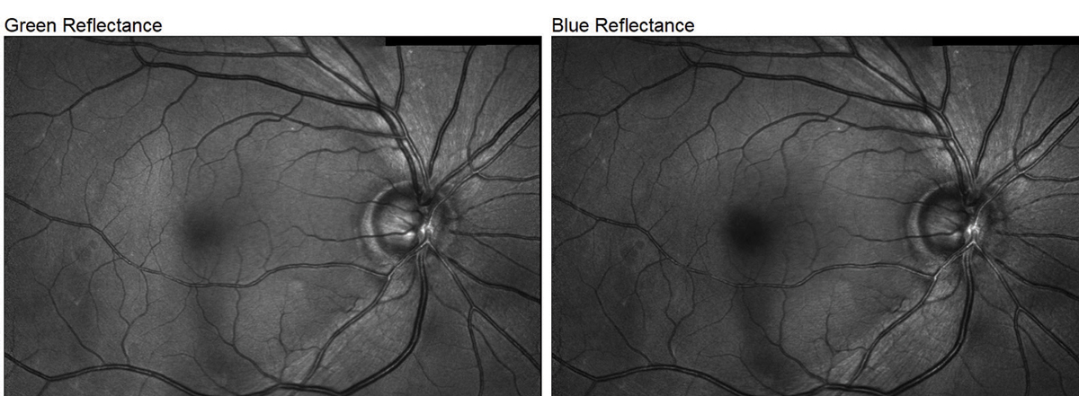
How Do OCT Devices for Glaucoma Compare?
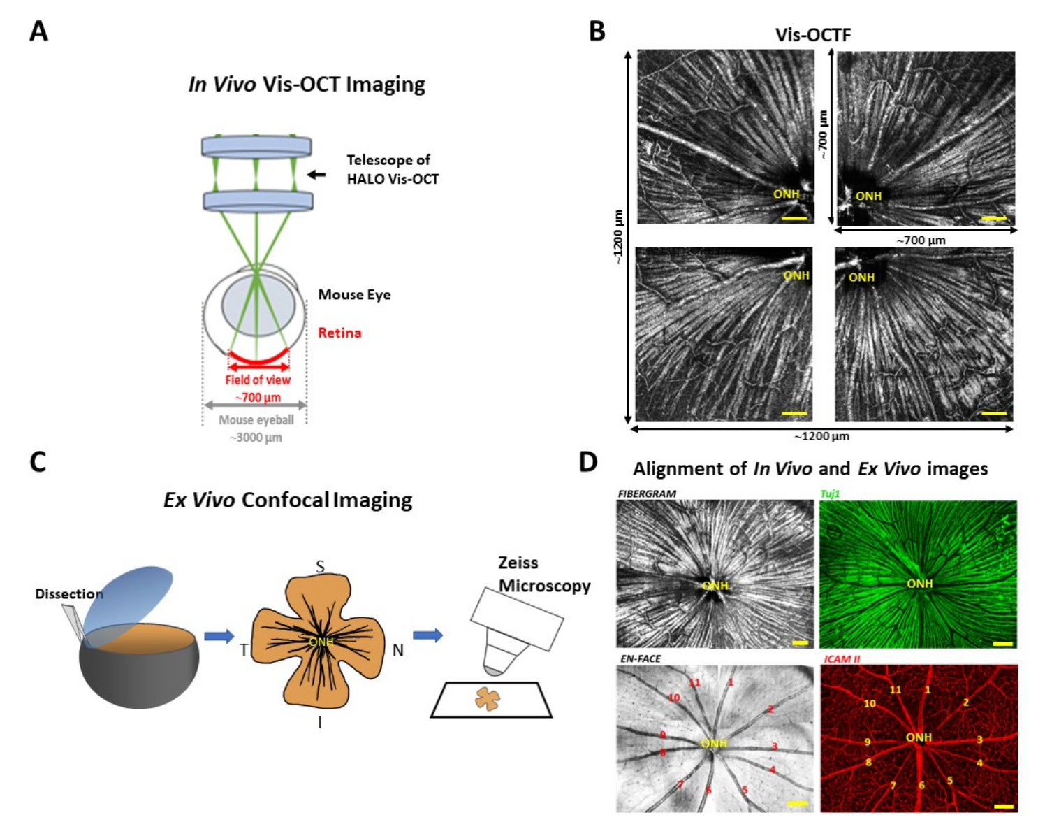
Alignment of Visible-Light Optical Coherence Tomography Fibergrams with Confocal Images of the Same Mouse Retina
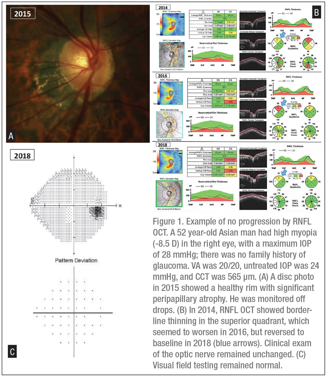
Monitoring Glaucoma Progression with OCT

En Face OCT Superior to Red-free Imaging for RNFL Defects

Parapapillary deep‐layer microvasculature dropout is only found near the retinal nerve fibre layer defect location in open‐angle glaucoma - Son - 2022 - Acta Ophthalmologica - Wiley Online Library
